Leg Muscle Diagram Posterior - Muscles of the hips and thighs | Human Anatomy and ... : Each of these muscles is a discrete organ constructed of skeletal muscle tissue, blood vessels, tendons, and nerves.
Leg Muscle Diagram Posterior - Muscles of the hips and thighs | Human Anatomy and ... : Each of these muscles is a discrete organ constructed of skeletal muscle tissue, blood vessels, tendons, and nerves.. They allow you to move and provide support for your upper body. A muscle along the outside of the leg that bends the foot out at the ankle. The posterior compartment of the leg is supplied by the tibial nerve. Dissection of right lateral cervical region diagram. It contains the plantar flexors:
Tibialis posterior originates on the proximal 2/3 of tibia and fibula and inserts onto the medial cuneiform and navicular. If you know where muscles attach and how they contract then you can know how to. Anatomy muscle 3d illustration 3d rendering adductor magnus anatomical arthritis back biceps femoris body buttocks calf muscle diagram female fitness gastrocnemius glutes gluteus maximus gracilis. Note that the posterior head of the adductor magnus inserts. Their large size is one of the most characteristic features of the muscular.
Tendons In Foot Diagram Diagram Of Lower Leg Muscles And ... from i.pinimg.com One muscle, the popliteus, acts only on the knee joint. The posterior compartment of the leg is one of the fascial compartments of the leg and is divided further into deep and superficial compartments. A muscle of the hip originating on the lateral surface of the ileum and inserted in the greater trochanter of the femur. Medially rotates leg when flexed. Muscles of posterior compartment of the leg. Each of these muscles is a discrete organ constructed of skeletal muscle tissue, blood vessels, tendons, and nerves. Get in touch with us today! Muscle anatomy leg muscles diagram muscle diagram nerves in leg.
The flexor hallucis longus muscle (fhl) is one of the three deep muscles of the posterior compartment of the leg that distally attaches to the plantar surface of the hallux (great or big toe).
The following life study male figure sitting on the floor, shows a male the following life study lower torso and legs in a frontal view, shows the lower torso of a male figure. The muscle forms the floor of the popliteal fossa the muscle fibers extend through the posterior compartment of the leg and converge to form a solid tendon that passes behind the distal end of the. Our muscles of the leg quizzes and labeled diagrams are the best way to consolidate your knowledge. Dissection of right lateral cervical region diagram. Those of the superficial group constitute a powerful muscular mass, forming the calf of the leg. Learn the origin/insertion, functions & exercises all three of the adductors originate from the pubis (pubic bone) and insert into the femur (thigh bones). Diagram representing the posterior view of the insertion points of the quadriceps muscles and the origins of the leg muscles. Note that the posterior head of the adductor magnus inserts. Get in touch with us today! Their large size is one of the most characteristic features of the muscular. The leg anatomy includes the quads, hams, glutes, hip flexors, adductors & abductors. Tibialis posterior originates on the proximal 2/3 of tibia and fibula and inserts onto the medial cuneiform and navicular. Anatomy muscle 3d illustration 3d rendering adductor magnus anatomical arthritis back biceps femoris body buttocks calf muscle diagram female fitness gastrocnemius glutes gluteus maximus gracilis.
Muscles of medial compartment of thigh. Their main function is contractibility. Start studying leg muscles (posterior view). Different types of muscles of arm diagram. Deltoid triceps brachii brachioradialis extensor carpi ulnaris extensor carpi digitorum.
Free Anatomy Quiz - The Muscles of the Leg (posterior) Image from www.free-anatomy-quiz.com A complete list of muscular system quizzes; Male muscular system, full anatomical body diagram with muscle scheme, vector illustration educational poster. Flexion, medial rotation and adduction at hip. We'll break down the anatomy and function of the upper leg, knee, lower leg, ankle, and foot. Human muscle system, the muscles of the human body that work the skeletal system, that are under broadly considered, human muscle—like the muscles of all vertebrates—is often divided into striated muscle, smooth muscle, and cardiac muscle. Muscle anatomy leg muscles diagram muscle diagram nerves in leg. Get in touch with us today! This muscle diagram is interactive:
Click on the name of a muscle for a page about that muscle (works for most labels).
Its action causes plantar flexion and inversion of. This is the largest of the three compartments of the thigh. Rotator cuff muscle with anatomical posterior and anterior view expample. Different types of muscles of arm diagram. Their large size is one of the most characteristic features of the muscular. Posterior view of human muscular system. Dissection of right lateral cervical region diagram. Have a product modelling and rendering project?. Anatomy muscle 3d illustration 3d rendering adductor magnus anatomical arthritis back biceps femoris body buttocks calf muscle diagram female fitness gastrocnemius glutes gluteus maximus gracilis. Each of these muscles is a discrete organ constructed of skeletal muscle tissue, blood vessels, tendons, and nerves. You'll learn about the muscles, bones, and other structures of each area of the leg. Female hip and leg muscles labeled posterior view, 3d rendering. A muscle of the hip originating on the lateral surface of the ileum and inserted in the greater trochanter of the femur.
Medially rotates leg when flexed. They allow you to move and provide support for your upper body. Dissection of right lateral cervical region diagram. Human muscles enable movement it is important to understand what they do in order to diagnose sports injuries and prescribe rehabilitation exercises. Muscles, connected to bones or internal organs and blood vessels, are in charge for movement.
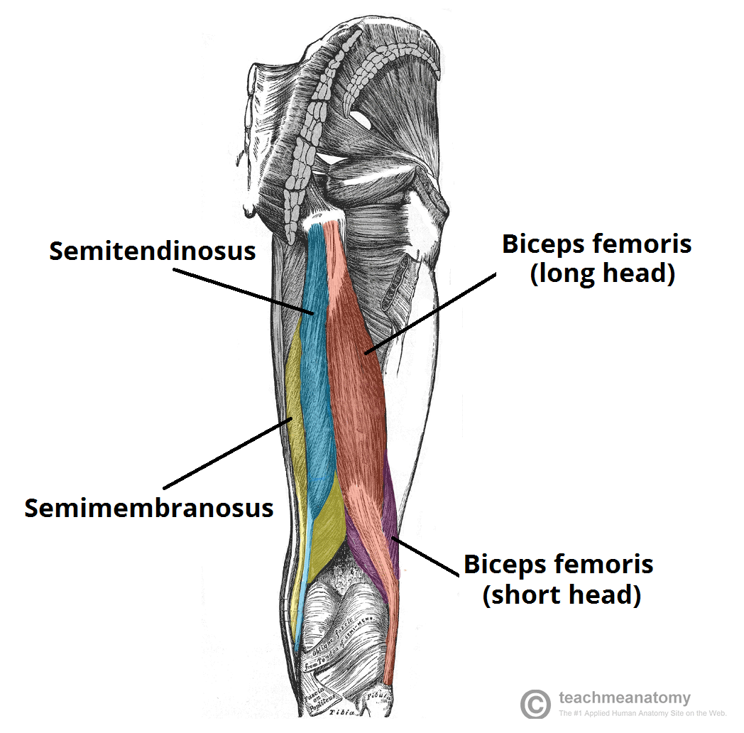
Muscles of the Posterior Thigh - Hamstrings - Damage ... from teachmeanatomy.info Covering upper limb, lower limb, head, back, and abdominal muscles through a series of muscular system quizzes. Your legs are two of your most important body parts. Human muscle system, the muscles of the human body that work the skeletal system, that are under broadly considered, human muscle—like the muscles of all vertebrates—is often divided into striated muscle, smooth muscle, and cardiac muscle. Posterior compartment, also known as the flexor compartment. Gastrocnemius muscle, large posterior muscle of the calf of the leg. One muscle, the popliteus, acts only on the knee joint. The large muscle of the posterior part of the lower leg. Muscles that move the leg.
3d medical illustration and rendering on leg posterior muscles for our client in australia.
Left leg, lateral (left) and posterior (right) views. This muscle diagram is interactive: Here we explain the major muscles of the human body. Medially rotates leg when flexed. Posterior muscles in the body. Gastrocnemius muscle, large posterior muscle of the calf of the leg. Muscles, connected to bones or internal organs and blood vessels, are in charge for movement. Note that the posterior head of the adductor magnus inserts. A complete list of muscular system quizzes; You'll learn about the muscles, bones, and other structures of each area of the leg. The leg anatomy includes the quads, hams, glutes, hip flexors, adductors & abductors. Learn the origin/insertion, functions & exercises all three of the adductors originate from the pubis (pubic bone) and insert into the femur (thigh bones). Anatomy muscle 3d illustration 3d rendering adductor magnus anatomical arthritis back biceps femoris body buttocks calf muscle diagram female fitness gastrocnemius glutes gluteus maximus gracilis.
Muscles of medial compartment of thigh leg muscle diagram. Everts foot at subtalar & transverse tarsal joints;